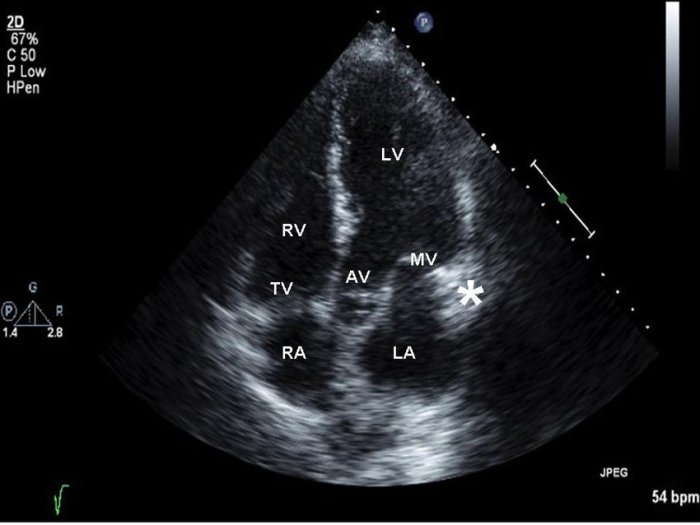
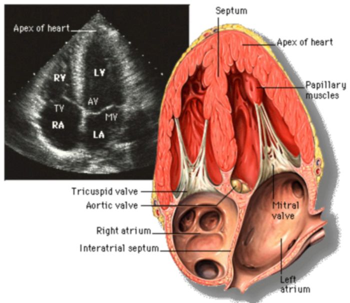
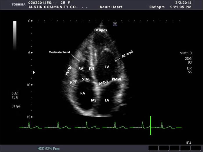
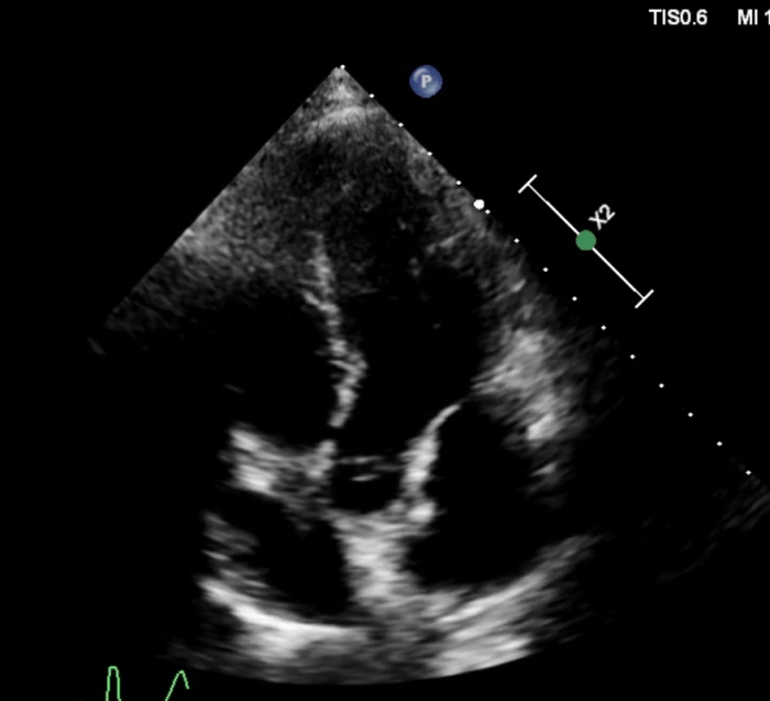
The apical 5 chamber view labeled provides a comprehensive visualization of the heart, allowing for a detailed assessment of its anatomy and function. This view is essential for diagnosing and managing a wide range of cardiac conditions.
This article will explore the apical 5 chamber view, including its anatomical landmarks, functional assessment, clinical applications, and advanced techniques. We will also provide answers to frequently asked questions about this important imaging modality.
Introduction to Apical 5 Chamber View

The apical 5 chamber view is a vital cardiac imaging technique that provides a comprehensive assessment of the heart’s structure and function. It offers a unique perspective, allowing clinicians to visualize the heart from the apex, or tip, of the left ventricle.
This view is essential for diagnosing and managing various cardiac conditions.
Imaging Modalities for Apical 5 Chamber View
Several imaging modalities can be used to obtain an apical 5 chamber view, including:
- Echocardiography:A non-invasive technique that uses ultrasound waves to create images of the heart. It is commonly used for obtaining apical 5 chamber views.
- Cardiac Magnetic Resonance Imaging (MRI):A non-invasive technique that uses magnetic fields and radio waves to produce detailed images of the heart. It can provide high-quality apical 5 chamber views.
- Computed Tomography (CT) Angiography:A minimally invasive technique that combines X-rays and contrast dye to create 3D images of the heart. It can provide detailed anatomical information but involves radiation exposure.
Anatomical Landmarks in the Apical 5 Chamber View

The apical 5 chamber view provides a comprehensive visualization of the heart’s chambers and major vessels. This view is crucial for evaluating cardiac anatomy, function, and any potential abnormalities.
The key anatomical landmarks visible in this view include the atria, ventricles, valves, and major vessels. Understanding the location, shape, and function of these structures is essential for accurate interpretation of the apical 5 chamber view.
Atria
- Right atrium (RA):The RA is located on the right side of the heart and receives blood from the superior and inferior vena cavae. It is a thin-walled chamber that helps fill the right ventricle.
- Left atrium (LA):The LA is located on the left side of the heart and receives blood from the pulmonary veins. It is a larger and thicker-walled chamber than the RA and plays a crucial role in filling the left ventricle.
Ventricles, Apical 5 chamber view labeled
- Right ventricle (RV):The RV is located on the right side of the heart and pumps blood to the pulmonary artery. It has a triangular shape and a thick muscular wall to generate enough force for pulmonary circulation.
- Left ventricle (LV):The LV is located on the left side of the heart and pumps blood to the aorta. It is a larger and more muscular chamber than the RV, reflecting its greater workload in systemic circulation.
Valves
- Tricuspid valve:The tricuspid valve is located between the RA and RV and prevents backflow of blood into the RA during ventricular contraction.
- Mitral valve:The mitral valve is located between the LA and LV and prevents backflow of blood into the LA during ventricular contraction.
- Pulmonary valve:The pulmonary valve is located between the RV and pulmonary artery and prevents backflow of blood into the RV during ventricular relaxation.
- Aortic valve:The aortic valve is located between the LV and aorta and prevents backflow of blood into the LV during ventricular relaxation.
Major Vessels
- Superior vena cava:The superior vena cava carries deoxygenated blood from the upper body to the RA.
- Inferior vena cava:The inferior vena cava carries deoxygenated blood from the lower body to the RA.
- Pulmonary artery:The pulmonary artery carries deoxygenated blood from the RV to the lungs for oxygenation.
- Aorta:The aorta is the largest artery in the body and carries oxygenated blood from the LV to the rest of the body.
Functional Assessment Using the Apical 5 Chamber View

The apical 5 chamber view is a valuable tool for assessing cardiac function, providing insights into the heart’s pumping ability and valve function.
For those who want to master the details of apical 5 chamber view labeled, it’s recommended to practice with the ap psych unit 9 practice test . The questions in this practice test are designed to cover all the key concepts related to apical 5 chamber view labeled, so you can be sure that you’re well-prepared for your exam.
Various parameters can be measured using this view, including ventricular volumes, ejection fraction, and valve function. These parameters help clinicians evaluate the heart’s performance and detect any abnormalities.
Ventricular Volumes and Ejection Fraction
- End-diastolic volume (EDV):The volume of blood in the ventricles at the end of diastole (ventricular filling).
- End-systolic volume (ESV):The volume of blood remaining in the ventricles at the end of systole (ventricular contraction).
- Stroke volume (SV):The difference between EDV and ESV, representing the volume of blood ejected from the ventricles per beat.
- Ejection fraction (EF):Calculated as (SV / EDV) x 100%, it represents the percentage of blood ejected from the ventricles during systole.
Valve Function
- Mitral valve:Assessed for regurgitation (backward flow) or stenosis (narrowing).
- Tricuspid valve:Evaluated for regurgitation or stenosis.
- Aortic valve:Inspected for stenosis or regurgitation.
- Pulmonary valve:Assessed for stenosis or regurgitation.
The apical 5 chamber view allows clinicians to visualize the heart’s chambers, valves, and blood flow patterns. By measuring ventricular volumes and assessing valve function, this view provides a comprehensive assessment of cardiac function, aiding in the diagnosis and management of various cardiovascular conditions.
Clinical Applications of the Apical 5 Chamber View
The apical 5 chamber view provides valuable insights into cardiac structure and function, making it particularly useful in diagnosing and managing various clinical conditions.
Cardiac Valvular Abnormalities
- Assessment of mitral valve prolapse: The apical 5 chamber view allows visualization of the mitral valve leaflets and can detect prolapse, a condition where the leaflets bulge into the left atrium during systole.
- Evaluation of aortic valve stenosis: The view provides a clear view of the aortic valve and can assess its opening and closing, helping diagnose aortic valve stenosis, a narrowing of the aortic valve opening.
Congenital Heart Defects
- Detection of atrial septal defects: The apical 5 chamber view can visualize the atrial septum and identify defects, allowing for early diagnosis and monitoring of congenital heart defects.
- Assessment of ventricular septal defects: The view helps visualize the ventricular septum and detect defects, aiding in the diagnosis and management of congenital heart conditions.
Cardiomyopathies
- Evaluation of left ventricular function: The apical 5 chamber view allows assessment of left ventricular size, shape, and contractility, helping diagnose and monitor cardiomyopathies, diseases that affect the heart muscle.
- Detection of right ventricular dysfunction: The view provides visualization of the right ventricle and can identify signs of dysfunction, aiding in the diagnosis and management of conditions like pulmonary hypertension.
Other Clinical Conditions
- Assessment of pericardial effusion: The apical 5 chamber view can detect the presence of fluid in the pericardial sac, aiding in the diagnosis of pericardial effusion, a condition where fluid accumulates around the heart.
- Evaluation of cardiac masses: The view can help visualize cardiac masses, such as tumors or thrombi, assisting in their detection and characterization.
Advanced Techniques and Future Directions

Advanced techniques have emerged to enhance the information obtained from the apical 5 chamber view. 3D echocardiography provides a three-dimensional reconstruction of the heart, allowing for a more detailed assessment of cardiac structures and function. Strain imaging, which measures the deformation of the heart muscle, can detect subtle changes in myocardial function that may not be apparent on conventional echocardiography.
Ongoing Research and Future Directions
Ongoing research is exploring the use of artificial intelligence (AI) and machine learning algorithms to analyze echocardiographic images, including the apical 5 chamber view. This has the potential to improve the accuracy and efficiency of echocardiographic interpretation. Additionally, research is investigating the use of the apical 5 chamber view in personalized medicine, where echocardiographic parameters are tailored to individual patient characteristics to optimize treatment strategies.
Potential Applications in Disease Prevention
The apical 5 chamber view can play a role in disease prevention by identifying individuals at risk for cardiovascular disease. By assessing cardiac structure and function, echocardiography can detect early signs of disease, such as left ventricular hypertrophy or diastolic dysfunction.
This information can be used to guide lifestyle modifications and medical interventions aimed at preventing the development of cardiovascular disease.
Question & Answer Hub: Apical 5 Chamber View Labeled
What is the apical 5 chamber view?
The apical 5 chamber view is an echocardiographic image that provides a comprehensive visualization of the heart’s chambers, valves, and major vessels.
How is the apical 5 chamber view obtained?
The apical 5 chamber view is obtained using transthoracic echocardiography, which involves placing an ultrasound probe on the chest to generate images of the heart.
What are the clinical applications of the apical 5 chamber view?
The apical 5 chamber view is used to diagnose and manage a wide range of cardiac conditions, including heart failure, valvular heart disease, and congenital heart defects.